
How HDC connects predictions to clinical evidence
Explore how HDC combines medical imaging and clinical language to classify retinal disease and support review of AI predictions.

On this page
Medical AI becomes more useful when its predictions connect to evidence a clinician can recognize. A disease label can help organize a screening queue. Knowing which visible signs support that label gives the person reviewing it something to examine, question, and discuss.
A study by Salik and colleagues in Scientific Reports1 explores an intriguing approach: connect retinal images with clinical language using hyperdimensional computing (HDC). Its central aim is to recognize retinal diseases across different datasets and scanners without repeatedly retraining the model. Bringing clinical vocabulary into that comparison also suggests an opportunity to make its predictions more inspectable.
Imagine an eye clinic that needs to review the morning’s scans. A useful AI assistant would help the team move from “the system flagged this image” to a more specific question: “Does the scan actually show the finding associated with this suggestion?” Let’s follow how such a review could work.
Give the scan something meaningful to match
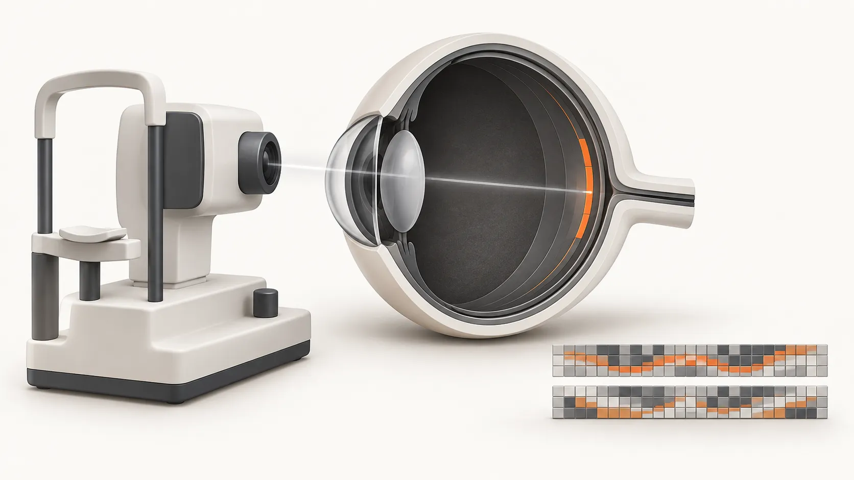
A technician uses an optical coherence tomography (OCT) scanner to capture a cross-section of the patient’s retina, the light-sensitive tissue at the back of the eye. The resulting image shows its layered structure. The clinician will examine it for signs of disease. Where could an AI assistant help?
The ML task is to classify the scan as showing diabetic macular edema (DME), age-related macular degeneration (AMD), or a healthy retina. In this approach, clinical descriptions guide both training and prediction. The model learns to match visual features with findings such as fluid associated with DME or deposits called drusen associated with AMD. For a new image, it predicts the disease category attached to the best-matching description.
The paper uses an image model called HDC+MLP. HDC provides the high-dimensional representation of visual information; the multilayer perceptron (MLP) supplies trainable neural-network transformations. A separate language model converts clinical descriptions into text vectors. During training, the image model’s weights are adjusted so its output aligns more closely with descriptions of the appropriate disease and less closely with alternatives. Clinical language therefore helps shape the visual features used for prediction.
The hypothetical AI assistant could show the matching description alongside the predicted category. If the match refers to fluid, the clinician has a specific finding to check in the original scan and assess in the context of the patient.
Two kinds of information, comparable patterns
A scan and a clinical description start as different kinds of data. HDC offers a way to bring their representations into a shared numerical space, where they can be compared. An encoder converts an input into a hypervector, a sequence of numerical values. HDC uses high-dimensional vectors called hypervectors, with information distributed across many values. In this application, training aligns the image and text representations so their similarity becomes useful for disease classification.
The authors’ implementation1 has two processing paths: the image encoder, HDC+MLP, converts the OCT scan into a visual representation; the text encoder, a language model, converts the clinical description into a text representation. The figure depicts each hypervector as a strip of shaded cells, with each cell standing for a numerical value. Similar patterns illustrate the agreement that training aims to produce between a scan and a relevant description.
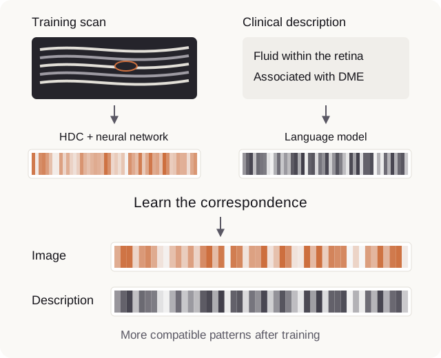
This alignment between image and text vectors is learned through contrastive training. The researchers first train the text model on clinician-reviewed disease descriptions. Its text vectors then guide updates to the HDC+MLP image model: a scan’s representation is pulled closer to descriptions of the corresponding disease and pushed farther from alternatives.
Repeating this across training scans teaches the model which visual and clinical patterns should agree. Our introduction to how HDC complements deep learning explores this division of work in more detail.
Here, the two sources remain separate for comparison. This is a multimodal application because it connects images and language. The text describes disease-related appearances, rather than an individual patient’s history. The researchers expanded clinician-written examples with AI-generated descriptions that clinicians reviewed.
Which description fits this scan?
The HDC+MLP encoder turns a new scan into a visual hypervector, which the system compares with the clinical text vectors. If the description linking retinal fluid to DME correlates most strongly, the system predicts DME, giving our proposed assistant a candidate finding to present for review.
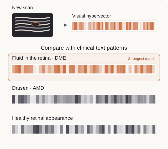
The paper evaluates disease predictions and image highlights. Building on those outputs, a proposed review screen could also expose the candidate descriptions used in the comparison:
| What the reviewer sees | What it helps them ask |
|---|---|
| The suggested disease and its matching description | Which finding made this suggestion relevant? |
| Other candidate descriptions and their scores | Was the choice clear, or do several interpretations compete? |
| The original scan with an image explanation | Is the highlighted evidence consistent with the proposed finding? |
Such a screen would make disagreement useful. A clinician could recognize a plausible finding but question the disease interpretation, or notice that an important alternative was never among the candidates. Keeping the description visible gives those objections a clear target.
Bring the comparison back to the image
Explainability gives the clinician a way to question a prediction using evidence they can inspect. If the system predicts DME because its strongest match describes retinal fluid, the next question is concrete: does the scan actually show fluid?
HDC+MLP supplies the visual hypervector used to select that description. To help locate supporting evidence, the paper also presents attention maps, overlays highlighting regions associated with the image model’s prediction. A review screen could show both, letting the clinician check whether the highlighted area supports the named finding.
The authors compared their highlights with expert-marked lesions in BIOMISA, one of the study’s retinal image datasets, but also reported missed tiny lesions. The clinician still needs to examine the rest of the scan. The practical benefit is a clearer basis for review: a disease suggestion connected to a named finding and visible evidence.
What could this enable for an eye clinic?
The researchers trained the model on retinal images from one public research dataset, known as the Zhang dataset. They evaluated it on held-out images from that collection and on three other public datasets without further training. The authors report stronger classification results than the comparison methods in their experiments. Live hospital testing remains future work, and the framework is limited to its trained disease categories.
For a clinic, the prospect is a screening assistant whose suggestions can be reviewed in the language staff already use. Applying it would require confidence that the descriptions cover the clinic’s cases and that useful performance carries over to its equipment and patients. A strong benchmark is a reason to investigate that fit.
HDC’s contribution here is its role in a learned image representation and explicit comparison with clinical text. The neural and language components contribute too. Image-language matching is a broader family of techniques; this paper offers a particular HDC-based way to build it.
The same question could guide other applications. A microscopy team might compare cell images with expert descriptions of morphology, provided those descriptions capture the distinctions being studied. A security team might compare event patterns with descriptions of known attacks, while keeping the underlying logs available for review. In both cases, the opportunity is to connect a machine’s match with evidence a practitioner can assess.
If you work with medical images, clinical data, or AI tools that support care teams, we’d love to hear where these ideas connect with your work. Perhaps you already have a model that makes useful predictions, but explaining those results to the people who rely on them remains hard. HDC could help make the evidence behind a prediction a more useful part of the clinical conversation.
Footnotes
-
Salik, A. Y., et al. Language assisted learnable hyperdimensional computing framework for retinal disease classification. Scientific Reports 16, 2892 (2026). Accompanying implementation. ↩ ↩2